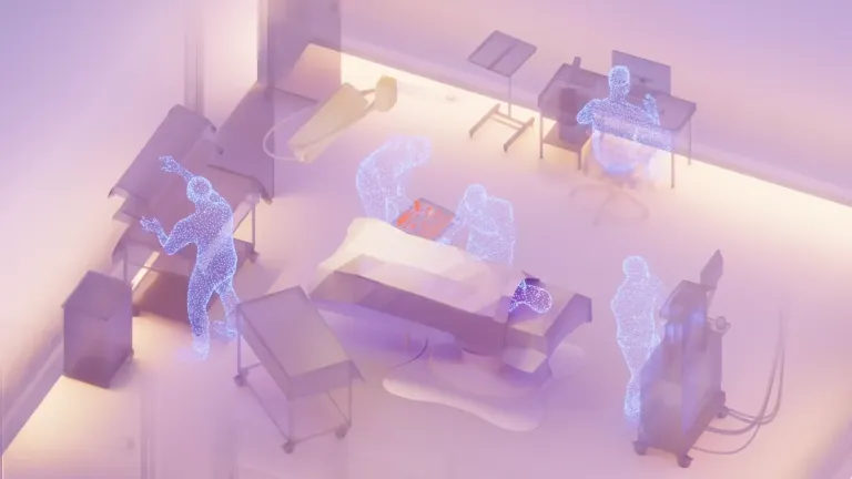
Why Operating Rooms Need 3D Spatial Intelligence.

Modern surgery is a spatial problem.
Surgeons operate within complex three-dimensional anatomy where millimeters matter. They navigate vessels, tissue planes, and instruments in confined spaces while coordinating with a multidisciplinary team. Yet much of the digital infrastructure surrounding the operating room still interprets the environment as if it were two dimensional.
The operating room is one of the most resource-intensive environments in healthcare. Since every minute carries both clinical and financial weight, improvements in how we understand and manage the surgical environment are significant. [1]
3D sensing is becoming a key part of that shift.
But 3D can mean different things. A stereoscopic display gives a surgeon depth inside the operative field. A room-scale spatial system must do more. It must understand where people, instruments, and equipment are located, how they relate to one another, and how those relationships change throughout a case. This article is about that second form of 3D: spatial intelligence for the entire operating room.
Surgery Has Always Been Spatial
Minimally invasive surgery introduced video-based visualization into the operating room, but traditionally it did so using 2D camera systems that project a 3D environment onto flat displays.
Surgeons compensate for this by relying on indirect cues such as instrument motion, shading, and anatomical familiarity to reconstruct depth perception. With experience, this mental reconstruction becomes intuitive, but the brain is still continuously inferring depth from incomplete visual information.
Spatial reasoning becomes easier when the visual system receives real depth information rather than having to infer it. Studies comparing 2D and 3D laparoscopic visualization have found improvements in task speed and reductions in performance errors when depth perception is restored. [2, 3]
Just as true 3D visualization helps the surgeon understand depth inside the body, 3D spatial perception helps machines understand depth, position, and movement across the entire operating room. In both cases, spatial context reduces the need to reconstruct a three-dimensional environment from flat images. For the surgeon, that improves visualization. For the system, it creates the foundation for reliable room-level understanding and automation.
The Operating Room Is a Spatial System
A surgical procedure is not just a list of steps. It is a spatially structured workflow.
Clinicians move through defined zones around the patient. Instruments are introduced and exchanged in both predictable zones and times. Equipment positioning changes across procedural phases. Critical events occur within spatial contexts.
At the same time, the room is not a fixed stage. Staff change position. Lights move. Displays are pulled closer to the table. Imaging equipment enters and leaves. Different surgeons arrange the same room differently, and the setup can continue changing after the procedure has started.
Experienced surgical teams understand this intuitively, but digital systems generally do not.
Most hospital software relies on manual documentation and discrete data entry to capture what occurs during surgery. Timestamps, instrument counts, and key procedural events are recorded manually because the systems themselves lack situational awareness of the operating room. They rely on human input to cover this gap, similarly to how 2D laparoscopic imaging relies on human interpolation to reconstruct the body in their head. This is fundamentally a sensing problem.
To understand surgical workflows automatically, a system must interpret not only what is visible but also where objects and people exist within the environment, how they interact, and how the state of the room changes over time.
That requires three-dimensional spatial and temporal context.
From Pixels to Context
Traditional computer vision systems interpret images as collections of pixels. This approach works for detecting objects but struggles to represent relationships between them in physical space.
Multi-view 3D perception adds critical signals:
- Depth estimation to determine distance between objects
- Spatiotemporal localization and memory of clinicians, instruments, and equipment
- Motion trajectories of hands and tools across time
- Relative positioning between participants in the procedure
These signals allow systems to move beyond recognition toward active understanding. All while keeping this understanding even if some cameras do not have a direct view, leading to unparalleled robustness
Recognizing a surgical instrument in an image is one relatively easy task that can be done with a single camera. Understanding that the instrument entered the operative field, interacted with tissue, and was passed back to a scrub nurse requires multi-view 3D spatial reasoning across time. Even more so when you consider that the room changes configuration during and between cases.
Three-dimensional perception across time enables that level of interpretation. When these signals are combined with advanced machine learning models, the operating room can be represented as a dynamic system with state changes on which automation can be built with confidence. Research in surgical data science and contextual AI is moving toward exactly this kind of context-aware understanding. [7, 8, 9]
Seeing the Whole Room: Why Coverage Matters
The operating room is crowded and dynamic. Surgeons, anesthesiologists, scrub nurses, circulating nurses, surgical technologists, residents, and equipment can all occupy the same space. Each moves, reaches, passes instruments, adjusts monitors, and repositions throughout the case.
Occlusion is therefore not an occasional technical problem. It is a constant feature of the operating room.
A single camera will inevitably be blocked. A clinician can obscure the instrument table. A monitor can create a permanent blind spot. Mobile imaging equipment can move across the camera’s line of sight. Adding depth to that single viewpoint does not solve the problem. Even a LiDAR or RGB-D sensor still measures only the surfaces visible from its position.
A small number of viewpoints can reduce these gaps, but it cannot eliminate them. Even three or four cameras may be blocked simultaneously when several clinicians surround the patient, equipment is repositioned, or the room is configured differently for a particular surgeon or procedure.
Those gaps matter. A sponge may be placed while every available view is blocked. An instrument may be passed behind a clinician. A relevant change in the sterile field may occur outside the covered area. If the system cannot observe an event, it cannot record it, understand it, or trigger a reliable response.
This changes how the sensing architecture should be designed. The goal should not be to install a predetermined number of cameras. It should be to provide enough well-positioned cameras and complementary sensors to achieve reliable, overlapping coverage of the room.
Critical areas should remain visible from multiple independent angles, even when staff and equipment move. If one viewpoint is blocked, several others should still be able to observe the same area. The correct number and placement of sensors will depend on the room layout, equipment, procedure types, and expected movement patterns.
This is where high-resolution 4K cameras become important. The operating room contains small instruments, subtle hand movements, device states, color, and other visual details that help distinguish one activity from another. A sufficiently distributed network of 4K cameras can preserve that detail across the room while providing the overlapping viewpoints needed to reconstruct spatial relationships and follow movement over time.
The operating room, like most of the physical world, was built for humans. And as humans, we understand their environment through visual cues. Color coding, labels, displays, indicator lights, packaging, gestures, uniforms, and equipment states all communicate information visually. We then combine these details with depth, movement, and prior context to understand what is happening around us.
This is another reason high-resolution cameras matter. They allow an AI system to observe the same visual layer of the environment that was designed to guide the people working within it. The system does not interpret the room exactly as a human does, but it can use many of the same visible cues.
The cost of neglecting this visual layer is not simply lower image quality. It can mean missing a critical cue—a color change, label, indicator light, gesture, or device state—that a member of the surgical team might otherwise recognize and act on. A geometry-only model may understand where something is without understanding what it is communicating.
A sufficiently distributed network of 4K cameras therefore provides more than coverage. It captures the human-readable context of the room from enough angles to remain useful when individual views are blocked.
LiDAR, RGB-D, and other sensors can strengthen this model by contributing direct depth, position, or device information. But they should complement the visual network rather than substitute for adequate coverage. Depth data cannot recover an event that every sensor was physically unable to observe.
The strongest direction is therefore a coverage-driven, multi-sensor architecture: enough high-resolution cameras to account for the room’s real occlusion patterns, combined with depth and other signals where they improve confidence. These inputs can then be fused into a continuously updated spatial and temporal model of the operating room.
The important question is not whether the system has one camera, four cameras, or a particular type of depth sensor. It is whether the architecture has been designed and validated so that critical activity remains observable as the room changes.
Operating-room research has repeatedly adopted multi-view and multimodal sensing because occlusion, clutter, and changing room configurations are fundamental characteristics of the environment, not rare exceptions. [4, 5, 6, 10]
This is not about adding cameras for the sake of collecting more video. It is about creating sufficient spatial redundancy for the system to be relied upon. Reliable observation enables reliable events, and reliable events are what allow hospitals to automate work and act in real time.
Why Spatial Context Enables Automation
Healthcare has invested heavily in digital systems, yet much of the operational context of surgery still depends on manual capture. Circulating nurses track procedural milestones. Staff document device usage. Surgical timelines are reconstructed after the case.
These tasks are necessary for compliance, quality tracking, billing, and patient safety. But they are fundamentally observational tasks.
If a system can perceive the surgical environment in three dimensions, continuously and across multiple viewpoints, many of those observations can be captured automatically.
Examples include:
- Detection of procedural phase transitions
- Tracking staff presence, positioning, and interactions
- Recognition of instrument and device usage
- Automated surgical timeline generation
- Context-aware intraoperative and postoperative documentation
- Decision support for retained-object recognition and surgical counts
- 4D scene reconstructions for training and review
The key requirement is reliable spatial understanding of the room. A hospital cannot build dependable workflows around a system that detects an event only when the room happens to be arranged correctly. If an event is missed because a view is blocked, a downstream system cannot know whether it did not happen or simply was not seen.
Real-time understanding changes the value of the information. A retrospective record can explain what happened. A reliable event delivered while the case is still underway can give the right people or systems an opportunity to respond.
A recognized phase transition can support coordination outside the room. An unexpectedly prolonged phase can become visible sooner. Equipment and turnover workflows can begin from observed context rather than waiting for a phone call or a manual update.
The point is not for software to make clinical decisions or replace established safety processes. It is to provide dependable context early enough for authorized people and systems to act, while removing observational work from staff who already have more important responsibilities.
For hospital IT teams, reliability must also be measurable. Events need timestamps, confidence, auditability, and clear behavior when the system is uncertain. Privacy, security, access control, and integration are not additions around the edge of the product. They are part of whether the system can be trusted at all.
The goal is not another dashboard that asks staff to interpret more data. It is to remove the need for manual observation and documentation in the first place.
The Future: Context-Aware Operating Rooms
Operating rooms are gradually evolving into sensor-rich environments where spatial perception, computer vision, and machine learning can work together to understand clinical workflows in real time.
Other industries have already gone through a similar transition. Robotics, manufacturing, retail and autonomous systems rely heavily on 3D sensing because machines must reason about the physical world rather than just process images.
The same principle applies in healthcare.
For hospitals, the useful output is not more video. What they need is reliable, structured context: what happened, where it happened, when it happened, and how confident the system is. That context can be connected to operational and clinical systems without asking staff to review hours of footage.
If a system can observe the operating room as a structured spatial environment, it can begin to understand how procedures unfold, how teams coordinate, and where operational inefficiencies occur.
That understanding enables automation not by adding more interfaces, but by giving existing hospital workflows dependable information without requiring a person in the room to capture it manually.
At VitVio, this is why multi-view 3D spatial perception is central to how we approach the operating room. Understanding surgical workflows requires systems that can interpret context, movement, and environment across the room and across time. Flat images and isolated detections are not enough.
The Next Phase of Digital Surgery
Robotic surgery platforms have already normalized stereoscopic visualization for surgeons. Many clinicians trained on these systems find it difficult to return to flat 2D views afterward.
Spatial perception improves not only visualization but also how humans and machines understand surgical environments. The next phase extends that awareness beyond the surgeon’s display and into the operating room itself; from there, the whole hospital.
When operating rooms become spatially observable environments, the procedural context that currently exists in human memory, handwritten notes, and after-the-fact documentation can begin to be captured automatically, reliably, and continuously in real-time.
The goal is not surveillance. It is to remove administrative friction so clinical teams can focus on the procedure and the patient.
3D sensing is more than a better visualization. Multi-view spatial intelligence is the foundation for context-aware operating rooms and for the next generation of automation built within them.
--
Written by Thomas Knox and Peter Rennert, PhD. July 30th, 2026
This is an extension of our recent article on Med Tech World: 3D spatial AI: The missing layer in the smart operating room
Have thoughts, questions, or a perspective worth sharing? I’d love to hear from you, reach out to Thomas Knox — Founder & CEO, VitVio.
--
Sources
- [1] Childers CP, Maggard-Gibbons M. Understanding Costs of Care in the Operating Room. JAMA Surgery. https://jamanetwork.com/journals/jamasurgery/fullarticle/2673385
- [2] Sørensen SMD et al. Three-dimensional versus two-dimensional vision in laparoscopy: a systematic review. Surgical Endoscopy. https://doi.org/10.1007/s00464-015-4189-7
- [3] Monnet E. Three-dimensional versus two-dimensional laparoscopy: What is the evidence? Veterinary Surgery. https://doi.org/10.1111/vsu.14329
- [4] Srivastav V et al. MVOR: A Multi-view RGB-D Operating Room Dataset for 2D and 3D Human Pose Estimation. https://arxiv.org/abs/1808.08180
- [5] Basiev K et al. Open surgery tool classification and hand utilization using a multi-camera system. International Journal of Computer Assisted Radiology and Surgery. https://doi.org/10.1007/s11548-022-02691-3
- [6] Özsoy E et al. Holistic OR domain modeling: a semantic scene graph approach. https://pmc.ncbi.nlm.nih.gov/articles/PMC11098880/
- [7] Maier-Hein L et al. Surgical data science for next-generation interventions. Nature Biomedical Engineering. https://doi.org/10.1038/s41551-017-0132-7
- [8] Padoy N. Machine and deep learning for workflow recognition during surgery. Minimally Invasive Therapy & Allied Technologies. https://doi.org/10.1080/13645706.2019.1584116
- [9] Vercauteren T et al. CAI4CAI: The Rise of Contextual Artificial Intelligence in Computer-Assisted Interventions. Proceedings of the IEEE. https://doi.org/10.1109/JPROC.2019.2946993
- [10] Özsoy E et al. MM-OR: A Large Multimodal Operating Room Dataset for Semantic Understanding of High-Intensity Surgical Environments. Proceedings of the IEEE/CVF Conference on Computer Vision and Pattern Recognition. https://openaccess.thecvf.com/content/CVPR2025/html/Ozsoy_MM-OR_A_Large_Multimodal_Operating_Room_Dataset_for_Semantic_Understanding_CVPR_2025_paper.html